Important note: This post is a discussion of one of the claims (claim in title) made in my book The Human Holographic Visual System. To learn more of how the retina and other components of the visual system function, read my book. It is available in hardback, paperback, and e-book formats at Amazon, Barnes and Noble, and Bookshop.
Because the light waves are nanoscale in size, the visual system processes described here are architecturally structured to operate at the nanoscale.
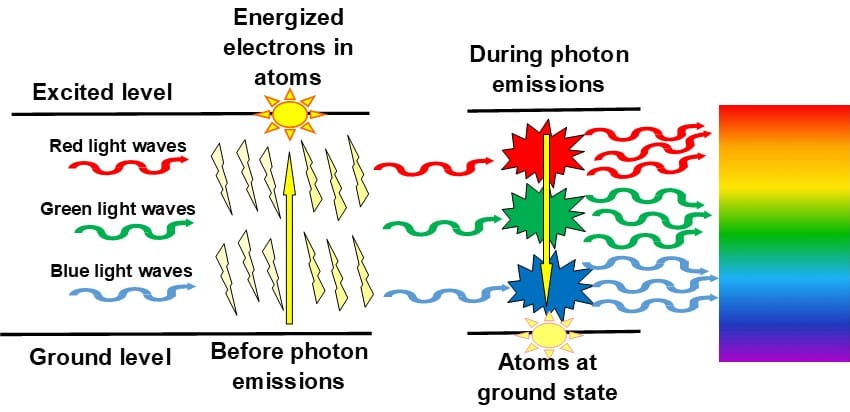
The light waves of the interference patterns leave Retinal Nerve Fiber Layer and flow through the layers of the retina on the outside of the neurons in the layers. When the light waves each the photoreceptors, the incoming light waves trigger the photoreceptors to flood the cell membrane with more positive sodium ions producing a metastable state for the cell membrane, especially the discs of the outer segment, with most of their molecules having their electrons energized.
Of the different types of opsin molecules available, the human visual system uses opsin1 for the rod and cone photoreceptors and further differentiates the opsin1 for the different receptors:
- Opsin1 rhodopsin for rod photoreceptors,
- Opsin1 OPN1SW for blue cone photoreceptors,
- Opsin1 OPN1MW for green cone photoreceptors, and
- Opsin1 OPN1LW for red cone photoreceptors.
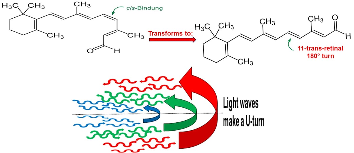
At the same time as the discs are being energized, the Opsin1 molecules with their 11-cis-retinal molecules in their pockets are capturing incoming light waves from the interference patterns.
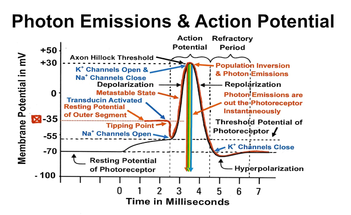
When the discs of the outer segment reach a peak of +30 mv, deactivation kicks in, collapsing the metastable state and dropping the voltage into the negative levels. At the same time, 11-cis-retinal molecules make a 180 degree turn and inject their captured original incoming light waves from interference patterns into the inside of the discs of the outer segment.
The reversal of polarity, population inversion, forces the energized electrons to release their excess energy in the form of a photon emission. And the simultaneous release of the original light waves from the 11-cis-retinal molecules strikes the light waves of the photon emission making it a stimulated photon emissions. Now, for each wavelength, all the light waves from the stimulated photon emission are identical in phase, frequency, and direction as the original incoming light waves from interference patterns. This means that:
- More of and only red wavelength light waves from the stimulated photon emissions are available for the red cone photoreceptors
- More of and only green wavelength light waves from the stimulated photon emissions are available for the green cone photoreceptors
- More of and only blue wavelength light waves from the stimulated photon emissions are available for the blue cone photoreceptors
- More of and only neutral-color wavelength light waves from the stimulated photon emissions are available for the neutral-color rod photoreceptors.
The discs of the outer segment act as microtubules that contain the stimulated emission and funnel the light waves through the inside microtubules to the inside of the inner segment of the photoreceptors. Because the light waves travel at the speed of light, the light waves of the interference patterns for each wavelength instantaneously rush out of the photoreceptors into the inside of the visual system but in the opposite direction. The light waves made a U-turn inside the discs of the outer segment.
To learn more, read my book The Human Holographic Visual System. The book is available in hardcover, paperback, and e-book formats at Amazon, Barnes and Nobles, and Bookshop.