Important note: This post is a discussion of one of the claims (claim in title) made in my book The Human Holographic Visual System. To learn more of how the retina and other components of the visual system function, read my book. It is available in hardback, paperback, and e-book formats at Amazon, Barnes and Noble, and Bookshop.
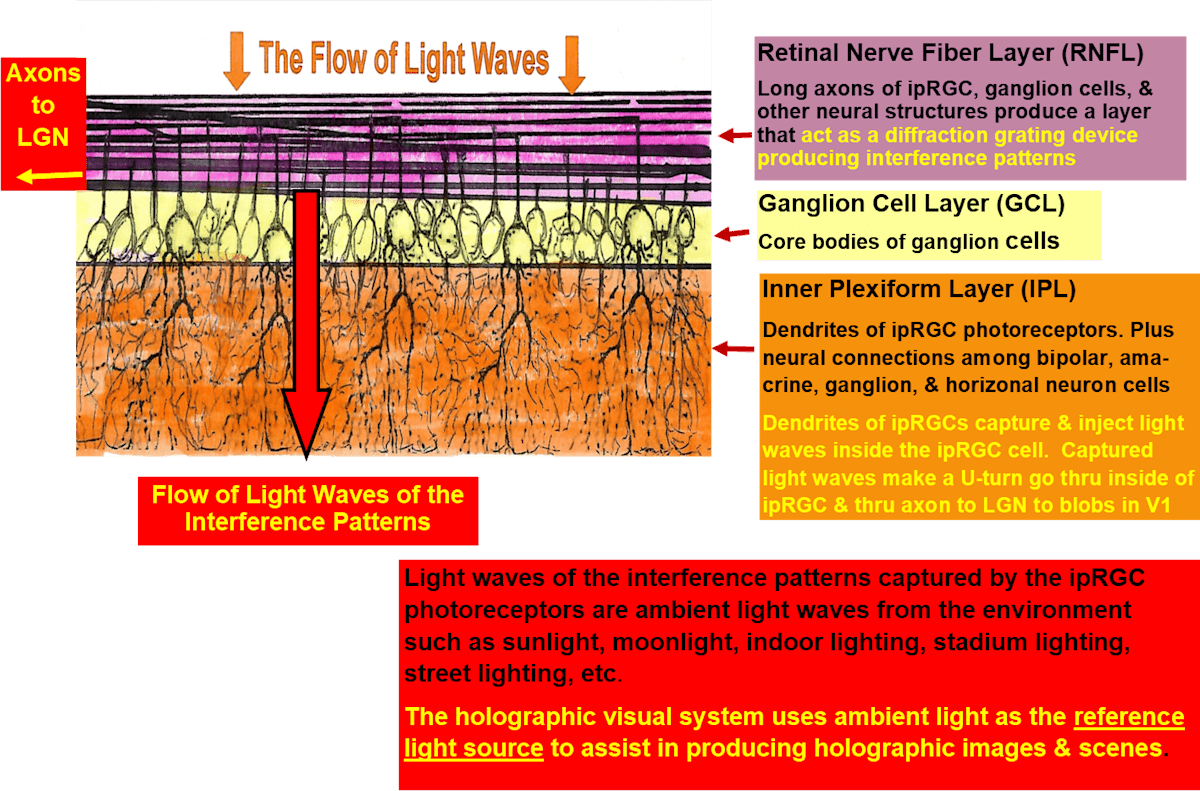
The ambient light waves from the immediate surrounding outside environment are the REFERENCE LIGHT SOURCE and are captured by the dendrites of the ipRGC photoreceptors.
After the Retinal Nerve Fiber Layer produce interference patterns, the light waves travel through a second layer called the Ganglion Cells Layer (GCL). Next, the light waves reach the layer called the Inner Plexiform Layer (IPL) which is the layer where the dendrites of the ipRGC photoreceptors exist.
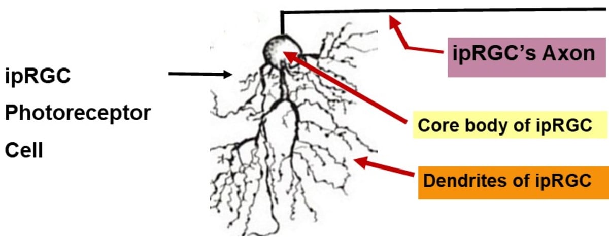
Like the rod and cone photoreceptors, the ipRGC photoreceptors use a type of opsin molecules (a photopigment) to capture light waves. This opsin molecule is opsin4 called melanopsin. The melanopsin (opsin4) resides on the plasma membrane of the ipRGC's dendrites, dendritic arbors, and cell body.
There are different types of ipRGC photoreceptors that range M1-ipRGC through M6-ipRGC subtypes. Also, there are different types of melanopsin that fit the different types of ipRGC photoreceptors, but they are hard to discuss in detail because there is little research literature related to them.
Like the other opsins in the photoreceptors, melanopsin molecules have a special ‘pocket’ where they hold a molecule of 11-cis-retinal. The 11-cis-retinal molecules capture light waves according to their length of conjugated double bonds. The ipRGC photoreceptors have one important difference than that of rod and cone photoreceptors, they do not use the population inversion and photon emissions processes.

An especially important process that the ipRGC photoreceptors have is that they are capable of signaling constant light continuously for at least 20 minutes. The ipRGC photoreceptors prolong their phototransduction which has the effect of blurring the spatial and temporal details within the interference patterns and leaving only the overall light intensity which is the ambient light (reference light source).
After capturing light waves, the 11-cis-retinal molecules (inside the pockets of melanopsin) rotate their molecular chain 180 degrees and inject their light waves inside the microtubules in the dendrites of the ipRGC photoreceptors. This action redirects the captured light waves in a reverse direction and through the inside of the ipRGC photoreceptors.
In the retina, the connections among the ipRGC photoreceptors, amacrine cells, and horizontal cells coordinate and synchronize the action potentials of the bipolar cells and ipRGC photoreceptors. Light waves of the ambient light are pulsed propagated with the action potentials through the axons of the ipRGC cells out the optic nerve and to the lateral geniculate nucleus (LGN). From the LGN, light waves of the ambient light are transferred to the “Blobs” in primary visual cortex.
To learn more, read my book The Human Holographic Visual System. The book is available in hardcover, paperback, and e-book formats at Amazon, Barnes and Nobles, and Bookshop.